How tumour testing decides which cancer drug you get

Not long ago, treatment was chosen by organ and stage: one set of drugs for lung cancer, another for bowel cancer. Today an oncologist increasingly looks not only at where the tumour grew, but at what is inside it — which broken genes are driving the cells to divide. There is only one way to find out: examine the tumour material itself.
For a number of modern drugs this is not an optional extra but a precondition: without a confirmed target, there is no basis to prescribe them. This article covers what sample is taken, which tests exist, what they actually decide and what they cannot do. It does not replace a consultation: decisions about testing and treatment are made by your treating oncologist.
Why the test comes before the drug
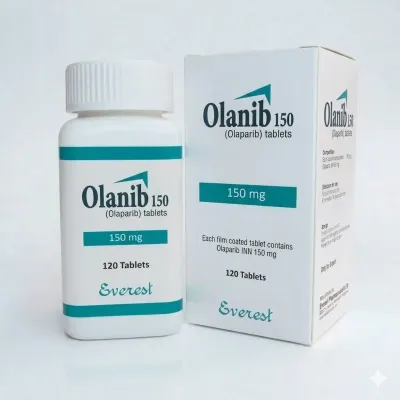
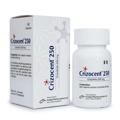
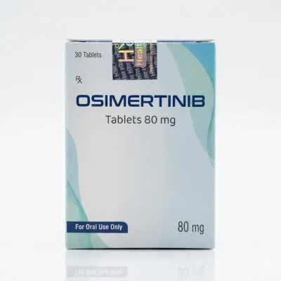
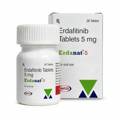
A targeted drug does not work against "cancer in general" — it works against one molecular target. Osimertinib shuts down a mutated EGFR receptor; with no mutation there is nothing to shut down. Olaparib kills cells that have lost the ability to repair double-strand DNA breaks, which is what happens with BRCA1/2 defects. Erdafitinib is used only when FGFR2 or FGFR3 alterations are present. Crizotinib and brigatinib only for ALK or ROS1 rearrangements.
So the logic is inverted: find the target first, then pick the drug. The test that confirms the target is called a companion diagnostic — it is paired with the drug and written into its label. Prescribing such a drug blind almost guarantees wasted months and money while the tumour keeps growing.
What sample is taken
There are only two sources, and they are not interchangeable.
Tumour tissue. The primary material — either a biopsy (needle, endoscope or during surgery), or an existing paraffin block left over from earlier surgery or diagnostic biopsy. If the block is stored in the pathology department, no new procedure is needed. It is worth finding out early where your block is and whether it can be released: this saves weeks and spares you a repeat procedure.
Blood. The so-called liquid biopsy: circulating tumour DNA (ctDNA) shed by dying tumour cells floats in the plasma. Collection is an ordinary blood draw, with no anaesthesia and no procedural risk. But the method comes with an important caveat, covered below.
Sample requirements people are rarely warned about
Molecular testing is not a matter of handing over any fragment. Laboratories have concrete requirements, and samples are often rejected as unsuitable.
- The specimen needs more than 20% tumour cells — if the section is mostly connective tissue or necrosis, the mutation simply will not be seen.
- Enough volume is required: typically around ten 5 µm sections and at least 50,000 cells to extract usable DNA.
- The material must be properly formalin-fixed and paraffin-embedded. Decalcified bone biopsies are often unusable: the acid destroys DNA.
- Old blocks degrade over time, so the freshest available material is preferred.
Small biopsies in advanced lung cancer are a particular problem: there is little tissue to begin with, and it must be split between histology, immunohistochemistry and molecular testing. To make it stretch, laboratories ask that each biopsy core be embedded in its own cassette rather than combined into a single block.
Four kinds of test and how they differ
"Mutation testing" is not one test but a family of methods that differ in cost, turnaround and the question they answer.
| Method | What it shows | When it is used |
|---|---|---|
| IHC (immunohistochemistry) | Presence and amount of a protein in cells | PD-L1 before immunotherapy, HER2, mismatch repair (MMR) protein status |
| FISH | Gene rearrangements and amplifications | Confirming HER2, ALK, ROS1 when IHC is borderline |
| PCR | One specific known mutation | Fast, cheap checks of hotspots: EGFR, KRAS, BRAF V600E |
| NGS panel | Tens to hundreds of genes in one run | Many possible targets, negative standard markers, or a clinical trial is being considered |
The choice is straightforward. With one or two obvious candidate targets, PCR or IHC is faster and cheaper. For molecularly heterogeneous tumours — non-small cell lung cancer, cholangiocarcinoma, cancer of unknown primary — a broad NGS panel up front beats spending scarce tissue on a series of single tests.
Turnaround differs by an order of magnitude: PCR takes days, NGS usually from four working days in laboratories with in-house sequencing up to about three weeks when material is sent to an external provider. For aggressive tumours that gap matters clinically.
Biomarker to drug: how it works in practice
A few examples where the test result directly drives the prescription. Drugs are listed to illustrate the target-to-treatment link, not as a recommendation.
| Finding in the tumour | Drug class | Examples in the catalogue |
|---|---|---|
| EGFR mutation (lung cancer) | EGFR inhibitors | Osimertinib, Osicent |
| ALK or ROS1 rearrangement | ALK/ROS1 inhibitors | Crizocent, LuciBriga |
| BRCA1/2 mutation | PARP inhibitors | Olanib |
| FGFR2/FGFR3 alterations | FGFR inhibitors | Erdanat, Erdafixen |
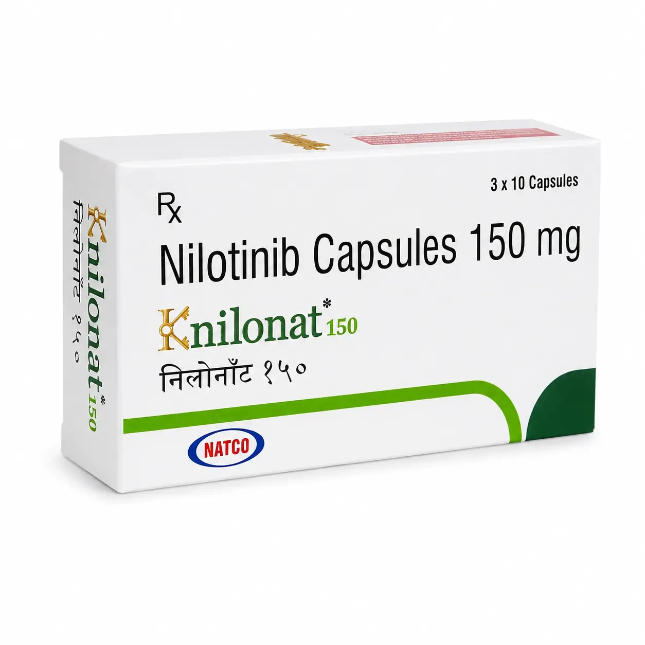
| BCR-ABL fusion (Ph+) | Tyrosine kinase inhibitors | Knilonat |
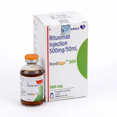
| CD20 protein on lymphocytes | Anti-CD20 antibodies | Reditux |
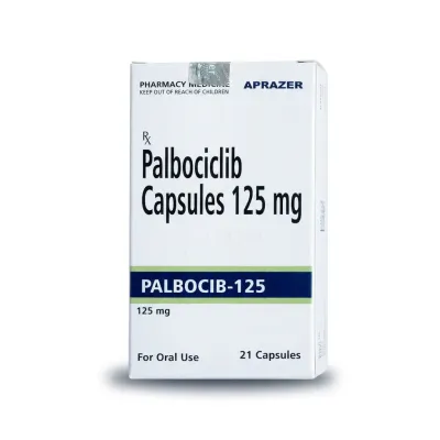
| Hormone-positive, HER2-negative breast cancer | CDK4/6 inhibitors | Palbocib, Abemaxen |
When the alteration matters more than the organ
The most unusual consequence of the molecular approach is drugs approved not for an organ but for an alteration, wherever the tumour grew. These are tissue-agnostic indications. There are nine of them, resting on six biomarkers: microsatellite instability (MSI-H) and mismatch repair deficiency, high tumour mutational burden (TMB), NTRK and RET gene fusions, the BRAF V600E mutation and HER2 overexpression.
How common is this? Across an analysis of more than 295,000 profiled samples, at least one such marker was present in roughly 21.5% of tumours — one patient in five. For someone with a rare cancer that has no standard of its own, this is sometimes the only route to a treatment that works.
Liquid biopsy: convenient, with one caveat
A blood test looks like the perfect substitute for biopsy: no anaesthesia, repeatable monthly to catch emerging resistance. In many situations that is exactly how it is used, especially when tissue is unavailable or the patient cannot tolerate another procedure.
But the method is asymmetric, and that matters. A positive result is almost always trustworthy: if the mutation was found in blood, it is there. A negative one often is not. Taken without stratification, agreement with tissue testing is modest: positive percent agreement around 63%, negative predictive value around 66%. In plain terms, roughly one in three "clean" blood results is wrong.
The reason is how much tumour DNA reached the plasma at all. Brain and intra-abdominal tumours shed poorly, so the assay simply does not see them. Modern reports therefore state the tumour fraction in the sample: below 1% a negative result is treated as uninformative and tissue confirmation is advised; at 1% or above, the negative predictive value exceeds 95%.
The practical takeaway: if blood finds nothing but the clinical picture still points to a target, that is a reason to go after tissue rather than close the question.
The molecular tumour board reads the report
An NGS report runs to dozens of pages of gene variants and does not treat anyone by itself. It is reviewed by a molecular tumour board, where an oncologist, pathologist, geneticist and clinical pharmacologist decide together which findings are clinically meaningful.
Real-world numbers are sobering and encouraging at once. In a large Belgian study a broad panel found clinically relevant alterations in 81% of patients, against 21% with small standard panels. The board issued treatment recommendations for 69% of patients — but only 23% actually received matched therapy. The gap has mundane explanations: the drug is not available in the country, it is unaffordable, the patient deteriorated, trial enrolment closed.
Where matched therapy did start, the difference in outcomes was substantial: median overall survival of 18.5 versus 9.1 months compared with patients treated outside the board's recommendations.
What this testing does not do
Two very different approaches are often blurred together in marketing.
Molecular profiling answers one question: does the tumour carry a target for which a drug exists. It is embedded in international guidelines and mandatory for dozens of medicines.
Chemosensitivity assays promise something else: grow the patient's tumour cells in a dish, expose them to various chemotherapy drugs and report which one "worked". The American Society of Clinical Oncology is unambiguous here — using such assays to select chemotherapy is not recommended outside a clinical trial. No prospective trial has shown that choosing chemotherapy this way extends survival, and US payers classify these assays as unproven. A clinic selling "personalised chemotherapy selection from your own cells" deserves hard questions.
Organoids are a separate case — miniature tumour replicas grown from the patient's own material. Same idea, modern technology, and 2025 results are encouraging: in ovarian and gastric cancer series, predictive accuracy reached 90% and above. But these remain research cohorts of tens of patients rather than routine care. For now organoids are a reason to look for a suitable clinical trial, not a paid service with a guarantee.
Availability in Ukraine
Molecular diagnostics is available in the country: NGS on Illumina platforms is offered by CSD LAB among others, and specialised centres operate in Kyiv. Cost is the main barrier — broad panels remain expensive, and wartime conditions constrain access further.
It is therefore worth actively looking for manufacturer-funded testing programmes; they exist and change over time. NGS testing of HRR genes for patients with metastatic castration-resistant prostate cancer, for example, was funded by the manufacturer and free of charge to the patient. Oncologists and patient organisations track the current programmes — ask them.
Questions to ask your oncologist
- Are there targets worth looking for in my diagnosis at all?
- Is the existing paraffin block enough, or is a new biopsy needed?
- Is a single targeted test sufficient, or is a broad panel better so tissue is not spent twice?
- How long will results take, and could the treatment plan change in the meantime?
- If the blood test finds nothing, will we confirm on tissue?
- Is there a manufacturer programme or clinical trial that covers the test?
In short
Molecular tumour testing is not a fashionable add-on but the gate to an entire class of drugs. The material is tissue — most often an existing paraffin block — or blood, and blood confirms a finding well while ruling one out poorly. Broad panels find clinically relevant alterations in most patients, yet far fewer actually receive matched therapy, usually for logistical rather than medical reasons. Selecting chemotherapy "from cells in a dish" is a different thing entirely and is not recommended in routine care.
This article is for information only. All decisions about diagnosis and treatment are made by your treating physician.